
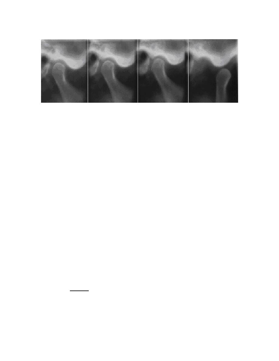
Figure 4-6. A series of radiographs of the temporomandibular joint produced by
tomography. The mouth is closed in the first three images and open
in the fourth.
4-16. TOMOGRAPHIC SYSTEMS
Radiography of a layer or section of the body is known by many names such as
tomography, stratigraphy, laminagraphy, and planigraphy. As a general rule, the names
were applied to the patented apparatus. However, the basic principles of operation are
the same. To standardize the nomenclature, the International Commission on
Radiological Units and Measurements (ICRU), in Handbook 89, recommends that
tomography be used to describe all body-section techniques. Handbook 89 further
classifies the systems according to the methods involved. This study guide describes
two moving tube systems, called rectilinear and pluridirectional.
a. Linear. The tube and film move in a straight line. This movement can be
described as plane-parallel. Some tomographic apparatus will only perform a linear
movement longitudinally to the long axis of the table. Others will perform crosswise and
diagonal movements with respects to the long axis of the table. See figures 4-7 and
4-8. Linear movements are often adequate for tomograms of the lungs, kidneys, and
simple bony structures.
b. Pluridirectional. Pluridirectional (multidirectional) systems, as a general
rule, produce better tomograms of areas that require maximum blurring, such as the
optic canal, inner ear, and complicated bone structures. In polytomography, the term is
usually used for pluridirectional system in which there is a wide variety of movements.
They include circular, elliptical, hypocycloidal, spiral, sinusoidal, and random
movements. The first three of these are discussed below.
(1) Circular. Although the circular movement is the simplest of the
polytomographic movements, it offers complete blurring. Circular movement is
accomplished with the tube tilted 20 to 40. The pattern is beneficial for views of the
lumbar spine.
MD0959
4-15

 Previous Page
