
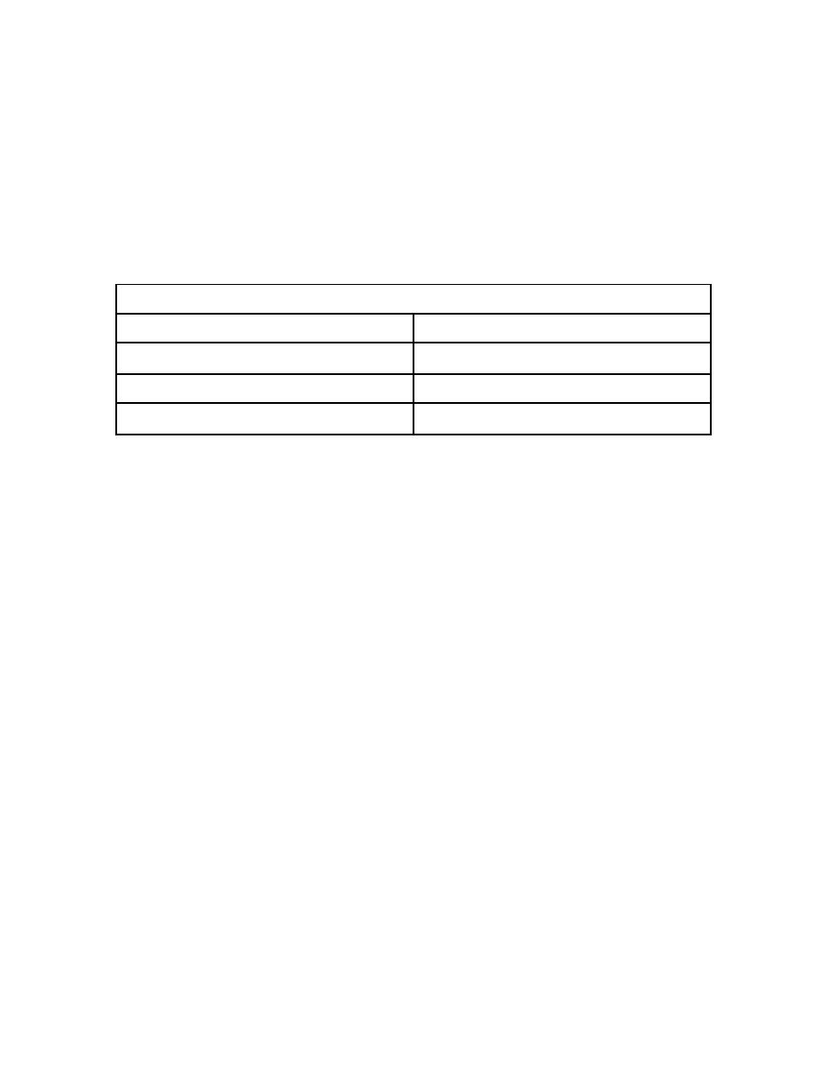
h. Technique factors. The technique adjustments in Table 4-1 are intended as
a starting guide only. Specific technique factors are listed for certain age groups and
fractional designations are given for other age groups. The fractional designations refer
to corresponding technique factors scaled for average adult patients. For example, "1/3
basic mAs" means that the technique factors customarily used for comparable
examinations of adult subjects remain constant, except that the mAs value is reduced to
one-third. This is not to imply that changing the mAs is the only way to adjust the
technique. The kVp, or both mAs and kVp, can be adjusted to provide satisfactory
results.
Pediatric Adjustments To Technique
0-1 year
Use 1/4 x mAs
2-4 years
Use 1/3 x mAs
5-11 years
Use 1/2 x mAs
12+ years
Use Adult technique
Table 4-1. Pediatric radiography: Suggested starting technique factors for
ages 0 to 12 years.
Section III. TOMOGRAPHY
4-15. INTRODUCTION
a. Since the introduction of computed tomography (CT) and magnetic resonance
imaging (MRI) with their excellent contrast resolution, tomography is less frequently
used. Conventional methods of radiography often resulted in the superimposition of
images on a two-dimensional film. To demonstrate a particular layer of the body
unobscured by images of overlying and underlying structures, a special technique
known as tomography can be employed. Tomography is now applied principally to
high-contrast procedures such as imaging the calcified stones in the soft tissues of the
kidney.
b. Tomography (body-section radiography) encompasses several methods by
which a specific layer of the body can be demonstrated to the exclusion of overlying and
underlying structures. Images of objects lying in other planes are eliminated, or at least
minimized, by blurring. For example, a lesion in the chest may be radiographed free of
overlying rib shadows. Figure 4-6 is an example of a tomogram of the
temporomandibular joint.
c. Tomography is a useful technique in all phases of radiography and can reveal
the correct diagnosis in many instances when it would otherwise be missed. It can be
used successfully with and without contrast media and plaster casts.
MD0959
4-14

 Previous Page
