
(4) A film is usually made to check the position of the catheter(s) and to
check patient position and technical factors.
(5) This film is immediately processed and presented to the radiologist or
the urologist for reading.
(6) At this stage of the procedure, the urologist will, in most instances,
retract the cystoscope, leaving the catheters in place.
(7) A contrast solution is introduced into the renal pelves and calyces
through the respective catheters by means of syringes. This procedure is accomplished
by the urologist.
(8)
At this point, the following routine is usually carried out:
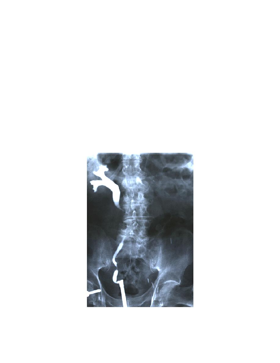
(a) At a signal from the urologist, the x-ray specialist exposes the
pyelogram (figure 2-31). For this exposure, the patient is instructed to suspend
respiration at the end of exhalation.
Figure 2-31. Retrograde pyelogram showing the urethral catheter in place and
the distribution of the contrast medium in the kidney and ureter.
MD0959
2-50

 Previous Page
