
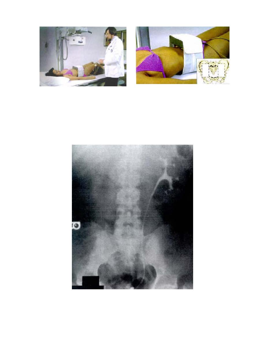
Figure 2-28. Patient has compression method applied.
(7) If subsequent pyelograms are to be taken, great care should be
exercised to match all films with respect to position, SID, radiographic contrast, and
density. This is necessary for satisfactory comparison with the results of prior
urographic examinations. A representative intravenous pyelogram is shown in figure
2-29. A labeled tracing of the pyelogram is shown in figure 2-30.
Figure 2-29. Intravenous pyelogram. Contrast medium in kidneys and ureters.
MD0959
2-47

 Previous Page
