
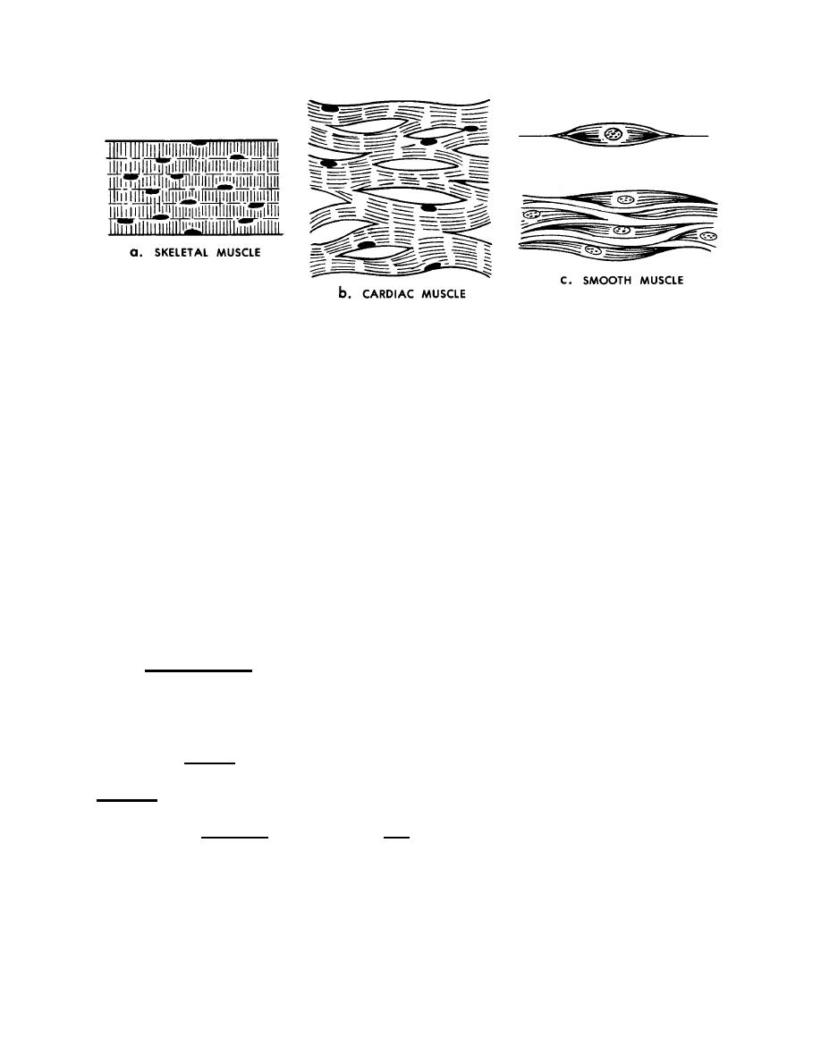
Figure 2-3. Types of muscle tissue.
b. Cardiac Muscle Tissue. The cells (muscle fibers) of cardiac muscle tissue
are short, branched, contain one nucleus, and are striated. This tissue makes up the
myocardium (wall) of the heart.
c. Smooth Muscle Tissue. The cells (muscle fibers) of smooth muscle tissue
are spindle-shaped, contain one nucleus, and are not striated. Smooth muscle tissue is
generally found in the walls of hollow organs such as the organs of the digestive and
respiratory systems, the blood vessels, the ureters, urinary bladder, urethra, and
reproductive ducts.
Section V. NERVOUS TISSUE
2-16. DEFINITION
Nervous tissue is a collection of cells that respond to stimuli and transmit
information.
2-17. NERVOUS TISSUE CELLS
a. A neuron (figure 2-4), or nerve cell, is the cell of the nervous tissue that
actually picks up and transmits a signal from one part of the body to another. A
synapse (figure 2-5) is the point at which a signal passes from one neuron to the next.
b. The neuroglia (also known as glia) is made up of the supporting cells of the
nervous system (glial cells).
c. The nervous tissues will be discussed in a later lesson.
MD0006
2-7

 Previous Page
