
1-3. INTERNAL FEMALE ORGANS
The internal organs of the female consists of the uterus, vagina, fallopian tubes,
and the ovaries (see figures 1-1 and 1-2).
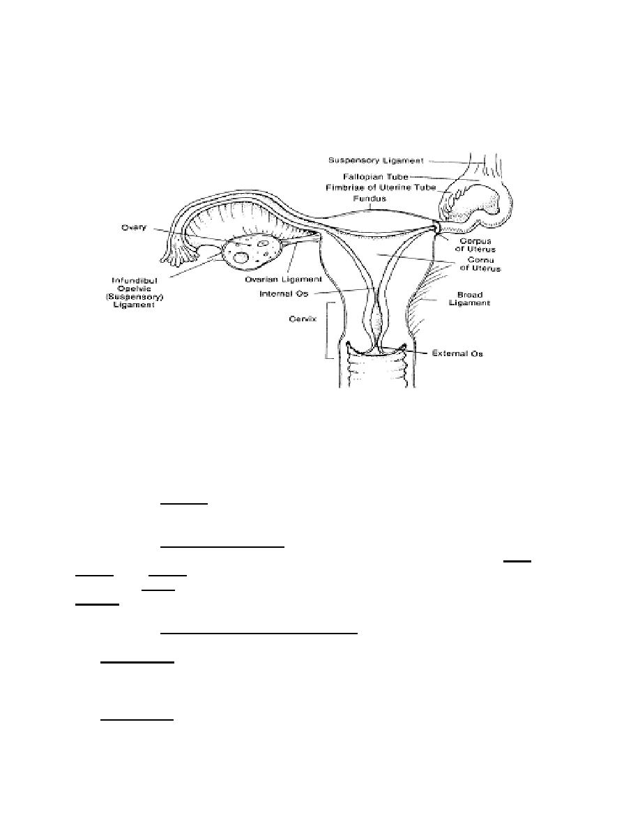
Figure 1-2. Anterior view of the uterus and related structures.
a. Uterus. The uterus is a hollow organ about the size and shape of a pear. It
serves two important functions: it is the organ of menstruation and during pregnancy it
receives the fertilized ovum, retains and nourishes it until it expels the fetus during
labor.
(1) Location. The uterus is located between the urinary bladder and the
rectum. It is suspended in the pelvis by broad ligaments.
(2) Divisions of the uterus. The uterus consists of the body or corpus,
fundus, cervix, and the isthmus. The major portion of the uterus is called the body or
corpus. The fundus is the superior, rounded region above the entrance of the fallopian
tubes. The cervix is the narrow, inferior outlet that protrudes into the vagina. The
isthmus is the slightly constricted portion that joins the corpus to the cervix.
(3) Walls of the uterus (see figure 1-3) . The walls are thick and are
composed of three layers: the endometrium, the myometrium, and the perimetrium.
The endometrium is the inner layer or mucosa. A fertilized egg burrows into the
endometrium (implantation) and resides there for the rest of its development. When the
female is not pregnant, the endometrial lining sloughs off about every 28 days in
response to changes in levels of hormones in the blood. This process is called menses.
The myometrium is the smooth muscle component of the wall. These smooth muscle
fibers are arranged. In longitudinal, circular, and spiral patterns, and are interlaced with
connective tissues. During the monthly female cycles and during pregnancy, these
MD0921
1-5

 Previous Page
