
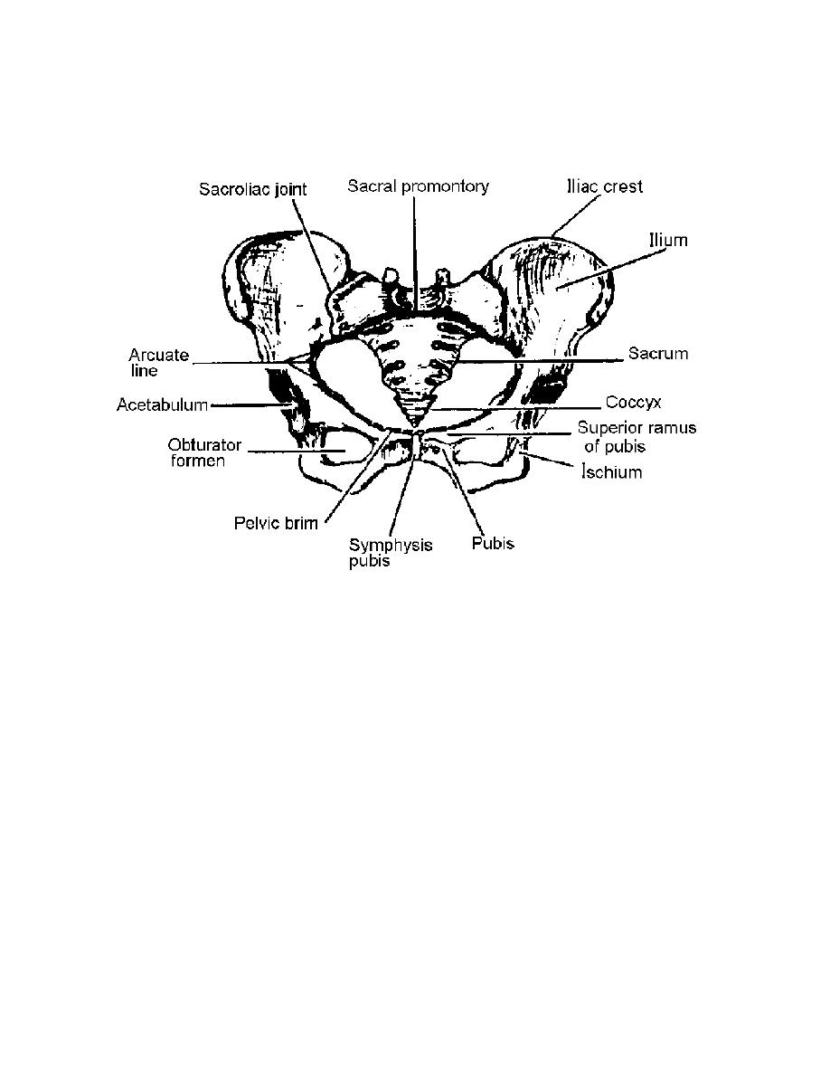
abdominal (false) portion from the true portion of the pelvis. The abdominal (false)
pelvis is the part above the arcuate line. The true pelvis is the part below this line. It
forms the passageway through which the infant passes during parturition.
Figure 2-2. The pelvic girdle.
b. The true pelvis may be considered as having three parts: the inlet, cavity, and
outlet. The muscles lining the pelvis facilitate movement of the thighs, give form to the
pelvic cavity, and provide firm elastic lining to the bony pelvic framework. All organs
located in the pelvis are covered by pelvic fascia. The fascia covering some muscles is
dense and firm, whereas that covering other organs is thin and elastic. The nerves,
blood vessels, and ureters passing through the anatomical structures are closely
associated with the muscular and fascial structures.
c. The pelvic fascia may be divided into three general groups: parietal,
diaphragmatic, and visceral. The parietal pelvic fascia covers the muscles of the true
pelvic wall and the perineum. The diaphragmatic fascia covers both sides of the pelvic
diaphragm, which is made up of the levator ani and coccygeal muscles. The visceral
fascia is thin flexible fascia that covers the pelvic organs. The floor of the pelvis, known
as the pelvic diaphragm, gives support to the abdominal pelvic viscera in this region.
The pelvic diaphragm, consisting of the levator ani and coccygeal muscles with their
respective fascial coverings, separates the pelvic cavity from the perineum. The basis
of modern vaginal surgery is concerned with the function of the levator ani muscles and
the provision of an effective lower outlet.
MD0928
2-3

 Previous Page
